

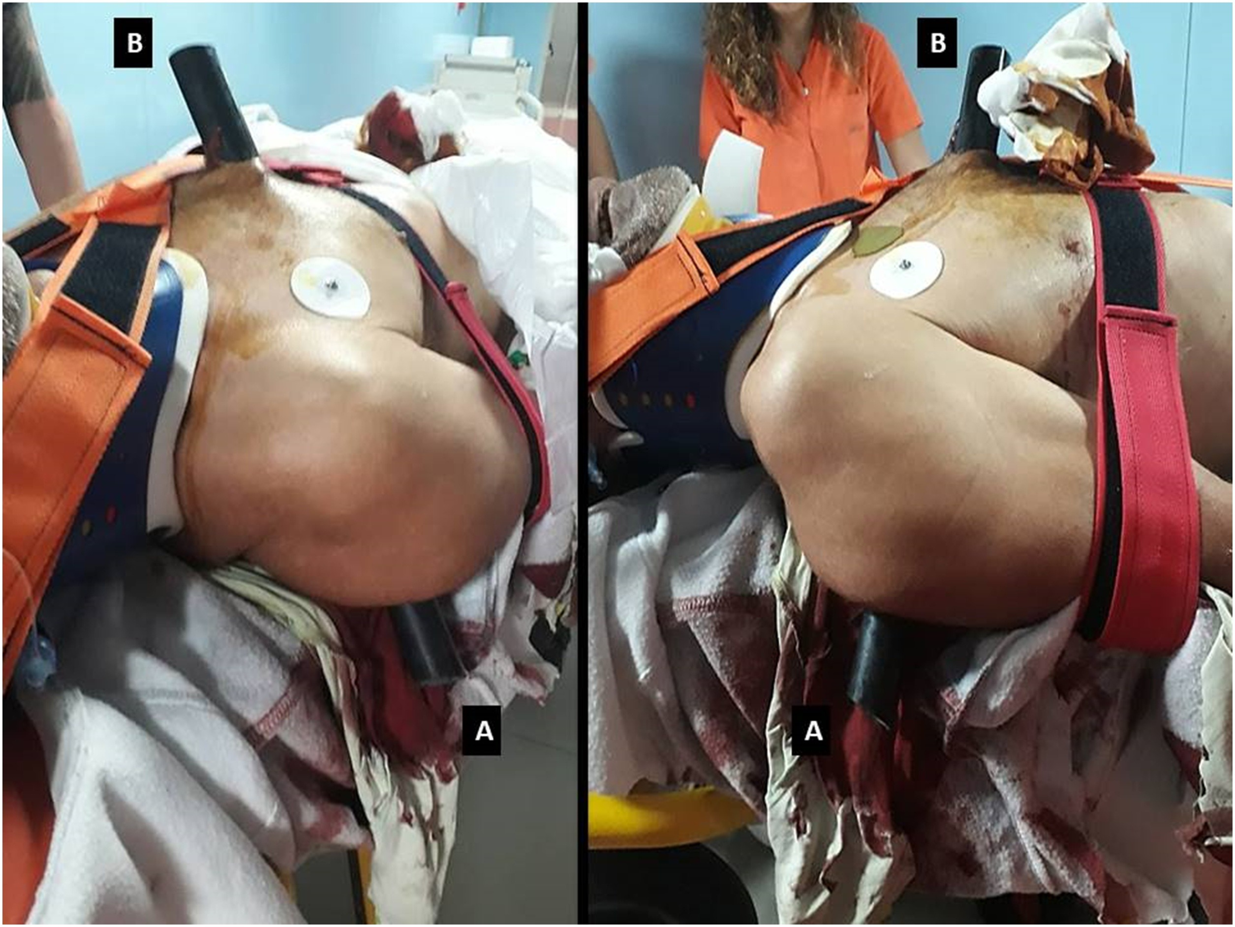
After approval from the ethics committee, we report the case of a 61-year-old male patient (61 kg, 168 cm, BMI 22) who by falling from a ladder impaled himself on a greenhouse pole. The pole entered from the right side of the back to exit from the sternum (Fig. 1).
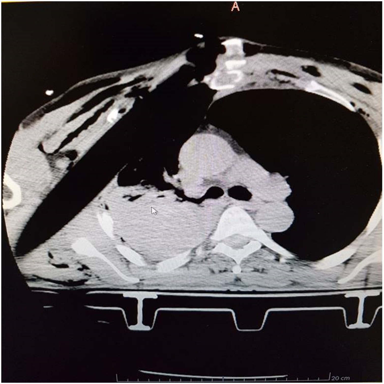
After prehospital trauma care, the patient was intubated and underwent a chest CT scan reporting “Tubuliform foreign body in the right lateral thoracic wall. Conspicuous apical-parietal-basal hemothorax with concomitant atelectasis. Front-basal right pneumothorax and decomposed fractal of the middle arch of the 3rd and 4th ribs” (Fig. 2). He was then referred to the oncologic thoracic surgery unit in our hospital.
On arrival the patient underwent a secondary survey: HR 100 bpm, NiBP90/60 mmHg, SpO2 100%, T 36.2 °C. Before surgery a further radiological study was performed: CT angiography excluded any involvement of the cardiac structures and of the larger vessels of the thorax. Total body CT was also performed.
Once in the operating room, intraoperative blood recovery was set up and a rapid intravenous infuser/warmer was used for fluid management. Large chemical heat pads were applied to the lower limbs. Endotracheal tube was removed after placing a bougie and a left Double Lumen Tube was inserted.
Waiting for radiologists to exclude any injuries to the spine and upper neck region, surgery started with the patient on spine board and with a cervical collar still on, which prevented from operating on the lateral position, which is the most suited.
A right anterolateral thoracotomy at 5th intercostal space was performed. After massive hemothorax (1200 ml) was drained, the intrathoracic part of the pole was clearly displayed. Hilar pulmonary structures, pericardium and epicardial structures were uninjured. A ventral subsegmentectomy of the right upper lobe was performed with parenchymal EndoGIA™(Medtronic. Minneapolis, Minnesota, USA) to remove a lacerated and contused lesion. After ligation of the intercostal arteries involved, the removal of multiple sternal and costal fragments allowed the exposure of the sheared internal mammary artery and vein: haemostasis with metal clips was performed.
A second right anterior thoracotomy at 3rd intercostal space, close to the thoracic intraparietal tract of the pole, was then performed to remove the foreign body. To prevent haemorrhage from the pole removal, a sterile gauze was fixed to a thoracic drainage, then the thoracic drainage tube was passed through the entrance hole of the pole (in the back) outside of the operating field. Then the removal of the foreign body started from the exit hole (in the parasternal area).In this way the gauze on one end acted as an haemostatic plug for the tissue injured by the pole penetration, exploiting its immediate mechanical haemostatic action. The pole was finally removed from the thorax (Fig. 3).
To guarantee stability and functionality of the rib cage despite the removal of the right 3rd and 4th ribs, the 2nd, 5th, and 6th right ribs were mobilized. Using a non-resorbable suture, the 2nd rib was pulled downwards and anchored to the lower ones, while the 5th and the 6th ribs were pulled upwards and anchored to the upper ones to fill the empty intercostal spaces.
Pectoralis muscle-myocutaneous flapping was performed and folded onto the contralateral pectoral fascia to cover the loss of thoracic wall. Two 28 Fr thoracic drainages were placed and a 16 Fr drainage was placed next to the entrance hole connected to a high-vacuum wound drainage systems.
After surgery the patient was referred to post-operative ICU. He was then successfully extubated. And on the 9th day a chest CT scan was performed reporting: “millimetric layer of pneumothorax, right basal pleural fluid layer, thickening band of the right upper lobe adjacent to metal suture and ground glass opacity residues from a likely outcome of alveolar haemorrhage”. On 12th day the patient underwent a spyrometric test: FVC 66% and FEV1 69% with a normal FEV1/FVC ratio as from mild restrictive pattern. Twenty days after surgery, the patient was dismissed. At one-month follow-up the patient underwent CT scan reporting no layer of pneumothorax or pleural fluid and the spyrometric test showed a mild improvement (FVC 76% and FEV1 79%). After three months a second follow-up was performed which confirmed the success of the intervention.
Management of chest impalement injury,
International Journal of Surgery Case Reports,
https://doi.org/10.1016/j.ijscr.2019.07.043.
(https://www.sciencedirect.com/science/article/pii/S2210261219304250)
Abstract: Presentation of case
We will describe the case of a man who impaled himself on a greenhouse pole by falling off a ladder.
Keywords: Chest wall; Trauma; Penetrating; Bleeding; Thoracic surgery