Lithopedion – A rare complication of abdominal pregnancy: A case report
Abstract
Dead foetal remains retained within the abdomen following an ectopic pregnancy leads to the formation of lithopedion, a rare entity. A 44-year-old woman sub-fertile for 16 years presented with sub-acute abdominal pain. She had not sought medical help earlier, on account of her religious or cultural beliefs. Imaging revealed a calcified mass containing bones. The differential diagnoses were lithopedion or a fetiform teratoma. During laparotomy, cutting open the mass revealed multiple long, short and flat bones, which together formed an almost complete foetal skeleton. Histopathology revealed an old ectopic gestation sac. This confirmed the diagnosis of lithopedion, estimated to be 32–34 weeks old at the time of foetal demise. This rare case highlights the impact of limited health literacy, along with strong religious and cultural influences, in delaying timely diagnosis and intervention.
Introduction
‘Lithopedion’ is a very rare complication of abdominal ectopic pregnancy, accounting for only 0.0054 % of all pregnancies [1]. It usually results from an undiagnosed and untreated advanced abdominal ectopic pregnancy [2]. It manifests as a calcified mass in the peritoneal cavity and can remain asymptomatic in the mother's body for years or present with gastrointestinal and/or genitourinary symptoms until it is discovered incidentally, following imaging or unrelated surgery [3].
This report concerns the case of a 44-year-old woman who presented with sub-acute abdominal pain and no reported history of pregnancy, who was found to have a lithopedion upon evaluation. This is apparently the first case of its kind to be reported from Sri Lanka.
2. Case Presentation
A 44-year-old woman who had been married for 16 years presented with a complaint of sub-acute pain in the lower abdomen and heavy menstrual bleeding for a few days. Despite having no reported history of conception, she had not sought medical attention for infertility due to her religious beliefs. She had been sub-fertile for 16 years and reported oligomenorrhoea. Her body mass index was 37.2 kg/m2. Physical examination revealed a palpable lump in the lower abdomen, hard in consistency and with restricted mobility.
Ultrasound examination revealed a heterogeneous mass in the lower right quadrant of the abdomen, with multiple hyper-dense and elongated areas within a poorly demarcated lower echoic lesion. Subsequent abdominal-pelvic X-ray in erect posture confirmed the presence of a radio-opaque mass containing hyperechoic bone-like structures in the right iliac fossa region (Fig. 1A). Her urinary and serum hCG were negative.
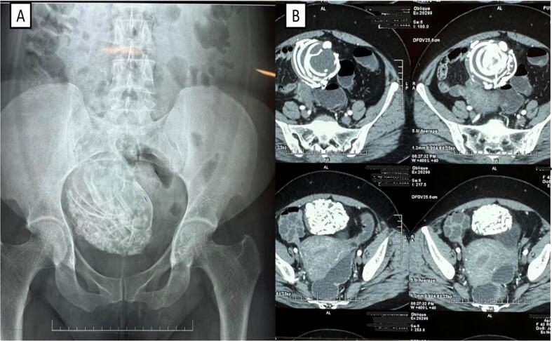 Imaging techniques used for diagnosis. An X-ray in the anteroposterior view (A) shows a calcified mass in the iliac fossa region. Contrast computed tomography (B) shows a dense mass measuring 9 × 7 × 7 cm with multiple bony foetal structures.
While the X-ray provided initial insights into the medical condition, further evaluation by advanced imaging modalities were deemed preferable to refine the differential diagnosis. Abdominopelvic-computed tomography (CT) with intravenous and oral contrast showed a heterogeneously dense mass measuring 9 × 7 × 7 cm placed to the right and anterior to the uterine fundus. It contained numerous formed bony elements, round and elongated. as well as curvilinear structures, representing long and flat bones. Central tissue elements measured 20 HU. No discrete placental mass was seen (Fig. 1B). Additionally, the CT scan revealed a closely placed bladder dome and few small bowel loops with loss of interphase. Lithopedion and fetiform teratoma were two differential diagnoses following the CT. However, the presence of axial skeleton bones usually excludes fetiform teratoma.
Based on these findings, a provisional diagnosis of lithopedion was made and exploratory laparotomy was planned. An infra-umbilical midline incision was made to have better surgical access. After entering the peritoneal cavity, an intra-peritoneal hard bony mass covered with a thick membrane was identified in the right adnexa. It was covered with some bowel loops and omentum. The mass received a blood supply from the omentum and the attached parietal peritoneum. The mass was excised following careful fine surgical dissections (Fig. 2A).
 Post-operative images of lithopedion. Following laparotomy, the calcified mass was removed (A). After the dissection of the mass, multiple bones were collected and assembled into a complete foetal skeleton (B).
An electric orthopedic bone saw was used to cut the outer thick membrane. After opening the mass, multiple long, short and flat bones were identified. It was possible to make an almost complete foetal skeleton by assembling bones that were inside the mass (Fig. 2B). The specimen was sent to a pathologist and routine abdominal closure was done. At the end of the procedure, cervical dilation and uterine curettage was performed. Post-operative recovery was uneventful and the patient was discharged from hospital 40 h after surgery.
Histopathological examination revealed an old ectopic gestation sac positioned within the mesosalpinx, encapsulated by a fibrous wall and omental tissue (Fig. 3).
Histopathological findings of the calcified mass. (A) Fibrous wall with bony spicules on the inner side and attached omentum on the outer surface. (B) Fibrous tissue on the inner side of the cyst wall. (C) The cyst wall attached to the fallopian tubal surface. The tube shows chronic salpingitis.
Remarkably, a well-formed foetal skeleton was identified within the sac. The degree of fibrosis and dystrophic calcification indicated that this was an old gestational sac. Additionally, the uterine curettage revealed degenerated endometrial changes associated with gestation. There was possible secondary infection in the degenerated decidua indicated by suppurative foci. By the length of the femur it was estimated that the lithopedion was 32–34 gestational weeks old at the time of fetal demise.
3. Discussion
Abdominal ectopic pregnancy is an uncommon type of extrauterine pregnancy; most cases occur after tubal rupture with a subsequent re-implantation in the peritoneal cavity. However, it can also occur when the zygote passes through the fallopian tube and is primarily implanted in the peritoneal cavity. These cases are sometimes not diagnosed and may resolve spontaneously, unless the foetus survives for more than three months as, before this time, the bones are still cartilaginous and the absorption will be fast and complete. If the dead foetus is too large to be absorbed by mother's body, the mother's immune system eventually recognizes the foetus as a foreign object and covers it with a calcium-rich substance that will eventually mummify and petrify the foetus to prevent infection [4].
Most cases of lithopedion remain asymptomatic for an extended period (4–60 years) [5] or present with chronic intermittent abdominal pain and gastrointestinal or genitourinary symptoms [1]. The absence of a reported history of pregnancy for the patient in the present case precluded determination of the exact duration of retention of the lithopedion. According to studies, the foetal demise in a majority (43 %) of cases of lithopedia occurs at term, followed by 7–8 months (27 %) and 3–6 months (20 %) of pregnancy [[6], [7], [8]]; in the present case, according to the femur length, foetal demise was at 32–34 gestational weeks.
Various conditions such as ovarian teratomas, uterine fibroids, calcified neoplasms, calcified aneurysms, inflammatory masses, dystrophic soft-tissue calcification and epiploic calcifications may present with a calcified abdominopelvic mass [9,10], and diagnosing a lithopedion can be challenging due to lack of specific clinical signs or symptoms [9]. In the present case, the non-specific appearance of the calcified mass necessitated imaging for a definitive diagnosis. Contrast CT confirmed a heterogeneously dense mass with the presence of axial skeletal bones, which was an indication to differentially diagnose lithopedion from fetiform teratoma. As the literature on this subject is mostly in the form of case reports, and clear evidence for the most appropriate therapeutic approach is lacking, laparotomy was performed, as reported in most of the similar cases in the literature. Moreover, the occurrence of complications even after several years makes surgical removal the best therapeutic choice for lithopedion, especially since the post-operative course is generally simple. No post-operative deaths have been reported [7].
Several studies have linked the occurrence of lithopedions to low socio-economic status and inadequate access to prenatal care due to poor health literacy and awareness [2,4,[9], [10], [11]]. Consequently, recent case reports predominantly emerge from regions with sub-standard medical services compared with developed countries [12]. Despite Sri Lanka's low socio-economic status, its provision of free and widespread access to healthcare has effectively prevented lithopedion and extrauterine pregnancy cases with severe clinical complications in the past. However, due to persistent issues such as limited health literacy together with strong religious beliefs and deep-rooted cultural practices in many parts of the country, individuals often delay seeking timely medical attention and, as a result, may not receive necessary healthcare. It is precisely such lapses in timely care and diagnosis that led to the development of lithopedion in the present case.
Increased access to and advancements in prenatal care and diagnostic techniques have led to a decline in lithopedion cases worldwide. By employing accurate diagnostic protocols, medical professionals can differentiate between ectopic and intrauterine pregnancies, thus mitigating the risk of acute or chronic complications [13]. Furthermore, enhancing reproductive and general health awareness among individuals in low- and middle-income countries, including Sri Lanka, can encourage utilization of healthcare facilities and services for their overall well-being. This approach holds significant potential for alleviating the health burden in these countries.
Citation: