
PLEASE, do not sad react on my facebook page, it's bad for the page.
Educational Medical Content
Only for Medical Students, view on your own risk!
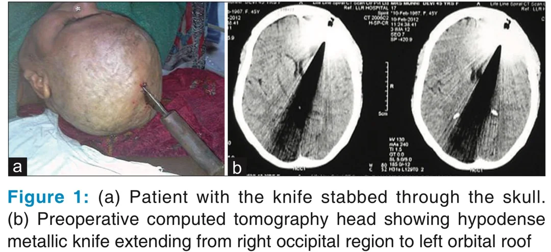
A female in her early forties was rushed by her relatives to our emergency department with a knife firmly embedded in her skull at vertex; with its wooden handle peeping out of it [Figure 1a]. Accompanying relatives told that she was found in such a state in her room about an hour back. According to the patient, she was suffering from repeated headaches for last 10 years. She stabbed this long knife with the help of brick into her skull to get rid of a headache. A rapid clinical evaluation revealed an anxious patient with preserved consciousness and orientation. She had pulse rate of 110/min, blood pressure 136/80 and respiratory rate of 24/min. Her Glasgow coma scale (GCS) was 15. Pupils were bilaterally normal size, reacting briskly to light with normal ocular movements. Her motor and sensory examination were normal. On local examination, there was a lacerated wound over vertex in right frontal region through which wooden handle and part of the knife were visible, there was no herniation of brain parenchyma from the wound. After ensuring hemodynamic stability, patient was taken for computed tomography (CT) scan of the head, which revealed a hypodense focus of metallic density (knife) extending from occipital and parietal lobes, deep brain structures, frontal lobe, reaching up to left orbital roof [Figure
1b].
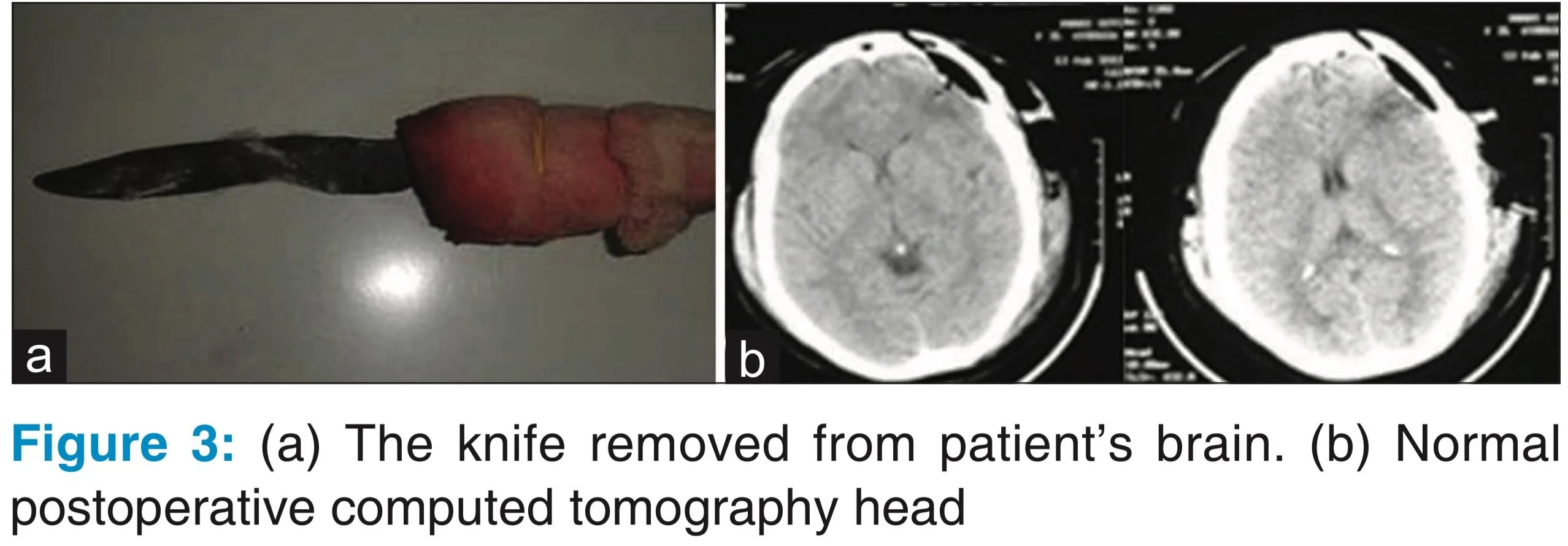
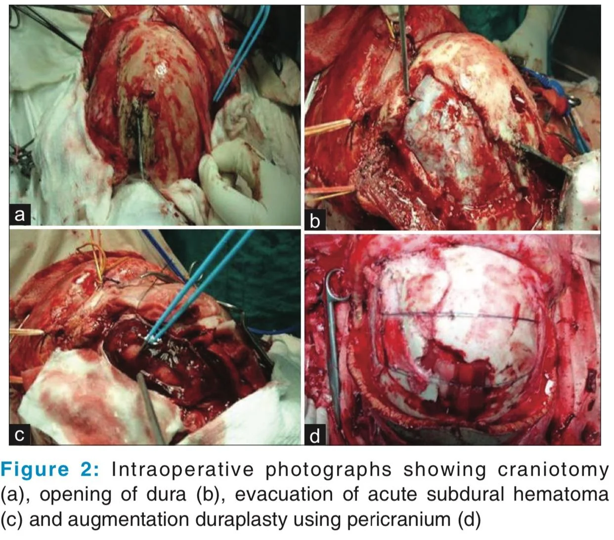
Patient was immediately shifted to emergency operation theatre after routine presurgical investigations. Left fronto-parietal craniotomy was made around the entry of knife and bone flap was removed in pieces holding the knife in position. On opening the dura, acute subdural hematoma (SDH) and contusion around the entry of knife was observed. The SDH was removed, and contusion was evacuated and the knife was slowly and carefully withdrawn. The tip of the knife was found traversing across the left frontal lobe and embedded itself in the roof of the left orbit. There were small bleeding points along the path of the knife which were controlled using bipolar coagulation. After satisfactory hemotasis, brain was lax and pulsating. Augmentation duraplasty was done using pericranium [Figure 2a-d]. The removed knife and postoperative CT scan of the head are shown in Figure 3a and b, respectively. Postoperative period was uneventful except superficial wound infection that was managed with dressing and antibiotics. Patient did not develop any neurodeficit during the recovery period. Postoperative CT scan did not show any residual or fresh hematoma.
Psychiatric and neurological evaluation of the patient was performed in the postoperative period, which revealed features of mixed anxiety and depressive disorder with migraine. Patient was put on antidepressant treatment, supportive psychotherapy and relaxation exercises along with migraine prophylaxis. Subsequently, patient was discharged on 10th postoperative day.