
FB removed the option to tap the links so the only way to easily find these articles is to tap the link in my Bio(About) section on the fb page.
Paywalls are everywhere. Knowledge shouldn’t be gated.
Your monthly coffee just makes them… less relevant. Much love to everyone supporting this concept.
A 36-year-old lady married for 20 years, Para 3 attended Gynaecology outpatient department with complaints of intermittent pain and swelling of lower abdomen over a period of one and half year. There was no history of any acute episode of pain or Missed periods. Her last childbirth was ten years back followed by puerperal tubal ligation. On clinical evaluation, there was a stony hard abdominal-pelvic mass reaching up to the umbilicus with restricted mobility in all directions.
The external genitalia was apparently normal. Systemic examination revealed no abnormality. Ultrasonography suggested the probability of a calcified subserosal fibroid. On laparotomy, pelvic structures were covered by omental adhesions, after dissection, a stony hard mass measuring 15×12 cms in right adnexa was visible. Mass was in close vicinity to the uterus and right ovary. Fallopian tube of same side could not be traced out. There were no ascites.
The rest of the abdominal cavity was normal (left tube and ovary were normal-looking) Differential diagnoses of ovarian teratoma, calcified leiomyoma of the uterus, lithopedion, and calcification of epiploic appendages kept in mind.
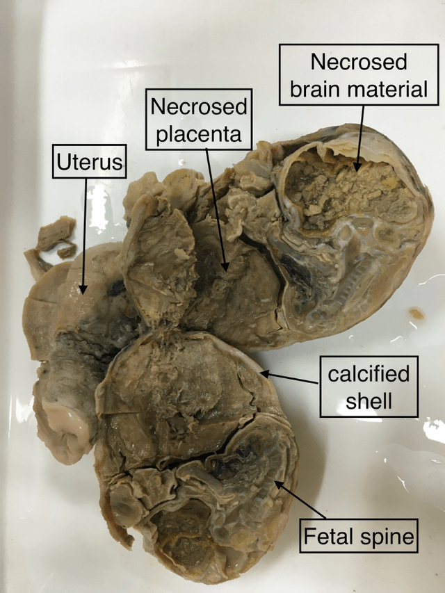 Fig 1: Gross image of the right adnexal mass showing macerated fetus with calcified shell
As patient belongs to a tribal community and was resident of an isolated Nicobar group of islands with low level of education and poor socioeconomic status with doubtful compliance to follow up. Moreover the mass was suspicious looking and patient had completed her family Decision for hysterectomy along with removal of right adnexal mass was undertaken. Patiently stood the procedure well with the uneventful postoperative period.
The specimen was sent histopathological examination. Cut surface of right adnexal mass revealed a macerated fetus with well-formed spine covered with calcified shell (Fig. 1). Microscopic examination also supported the same. Consequently, the Lithopedion diagnosis was supported radiologically, which reveals fetal skull, spines, ribs, and tubular bones (Fig. 2).
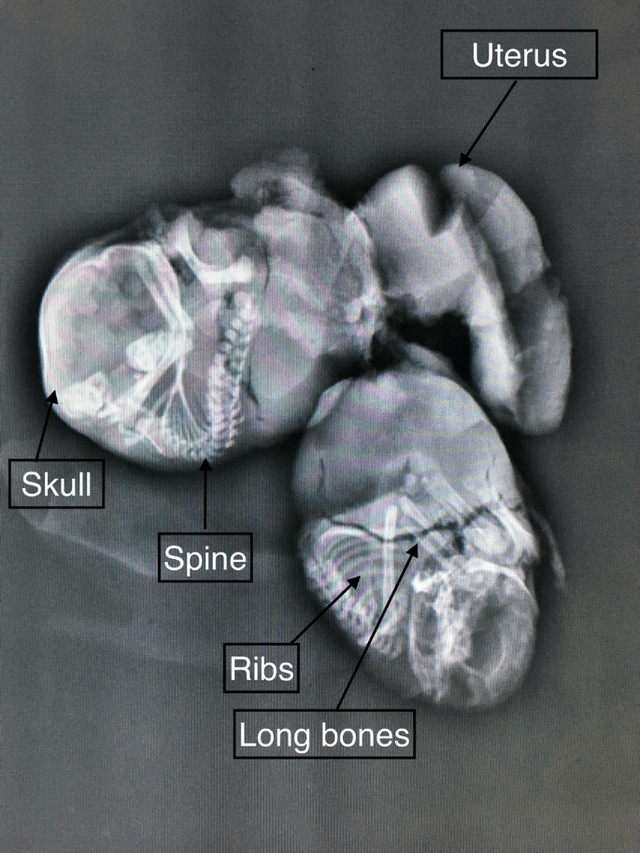
Fig 2: Plain X-ray showing fetal skull, well-formed spine, ribs, and long bones
Yadav, S., Sahoo, P.S.K., Choudhury, S. et al. A Perplexing Case of Lithopedion Masquerading as Subserosal Fibroid. J Obstet Gynecol India 72 (Suppl 2), 429–431 (2022). https://doi.org/10.1007/s13224-022-01620-3