

In October 2018, the police discovered a 45-year-old male deceased in the hilly areas of the district Sonbhadra, Uttar Pradesh, India. A preliminary postmortem was conducted there, but they could not conclude the cause of death. Four days past the first autopsy, the police then sent the body to our department at the Institute of Medical Sciences, Banaras Hindu University, Varanasi, for a re-postmortem examination.
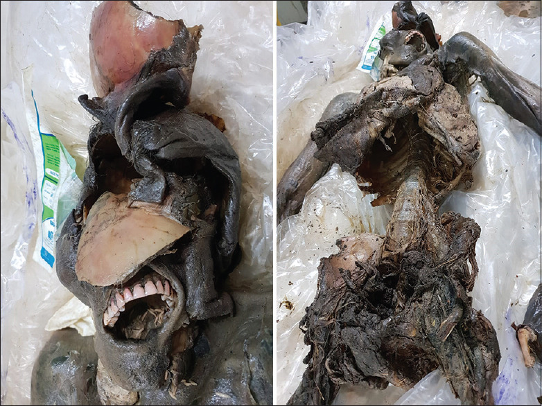
We received a decomposing body of a male with the scalp avulsed and the skull fragmented with brain matter missing. The skin is rubbery and darkens to a brownish-black color throughout the body. Maggot larvae are present in the mandibular, neck, and pelvic region and within the oral cavity. In addition, the anterior and posterior abdominal walls are missing, and the viscera are found liquefied [Figure 1]. A blown-out fracture of the skull is present, combined with laceration of the overlying skin, resulted in a large and continuous defect, extending from the left parietal and temporal region to the right parietal and temporal region, involving the frontal area and both orbits in the process. The breach is approximately 10 cm × 6.5 cm in size. The lower margin is in contact with the nose's root, while the upper margin is in touch with the vertex. The right margin is 3 cm from the pinna of the right ear, and the left margin is 5 cm above the left tragus [Figure 1]. The postmortem interval is estimated to be between 7 and 14 days. We retained the body for skeletonization to examine bony injuries in greater detail. After the skeletonization process, we reconfigured the skull and analyzed the bone for detailed damages.
The 12 pieces of the skull bones are present. They are numbered and manually reconstructed from these pieces. Following reconstruction, the following areas are identified as bone deficient [Figure 2]:
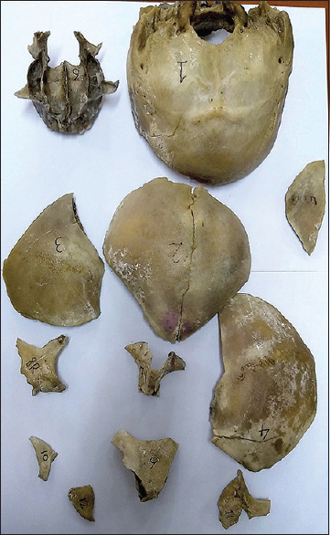
A portion of the left temporal, parietal, frontal, zygomatic, and maxillary bones are missing, resulting in a defect measuring 12.4 cm × 7 cm on the left side of the skull
2. A portion of the frontal bone measuring 7.5 cm × 3.7 cm along and to the left of the midline is missing
3. A portion of the right temporal and parietal bone is missing, resulting in a defect on the right side of the skull measuring 11 cm × 5 cm
4. On the left side, a portion of the sphenoid and the orbital bones are missing, resulting in a 3 cm × 4.5 cm defect in the middle and anterior cranial fossa
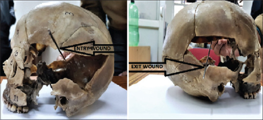
5. Furthermore, upon careful examination of the skull, a bullet entry wound is discovered on the left side's upper border of the defect, 7 cm above the tip of the left mastoid process and 6 cm posterior to the left orbital margin. The entry wound is shaped like an arc (forming the upper 1/3rd of a circle), with a smooth outer table and beveled inner table. In addition, a fracture line is observed originating from the entrance wound and extending backward between pieces 3 and 5. On the right side, the exit wound is present at the lower border of the defect. It is shaped like an arc (representing the lower 1/3rd of a circle), with a smooth inner table and beveling on the outer table. It is positioned 4 cm upward and back from the tip of the right mastoid process and 8 cm posterior to the right orbital margin. In addition, a fracture line is visible in piece number 1 that originates from the exit wound and extends backward and downward. After aligning the entry and exit with a probe [Figure 3], it is evident that the bullet traveled backward and downward from left to right.
Gupta M, Tiwari PK. An Autopsy Case of the Decomposed Body with Kronlein's Shot and the Importance of Skull Reconstruction. Int J Appl Basic Med Res. 2022 Jan-Mar12(1):47-50. doi: 10.4103/ijabmr.ijabmr_354_21. Epub 2022 Jan 31. PMID: 35265481; PMCID: PMC8848563.