

Mucormycosis Necrotizing Soft Tissue Infection: A Case Report of Fungal Infection Following a High-Speed Motorcycle Accident
Medical educational content for educational purposes only. Intended for medical students. Viewer discretion advised; continued viewing is at the viewer’s own discretion.
A 54-year-old male with a history of type 2 diabetes mellitus was initially evaluated in the trauma bay after a high-speed collision with a motor vehicle as the helmeted driver of a motorcycle. The patient was reportedly found pinned underneath a car. On arrival, the patient’s primary survey was significant only for diminished right anterior tibialis pulse and absent left anterior tibialis pulse, with no abnormalities of airway or breathing and a Glasgow Coma Scale of 15. Preliminary X-ray imaging revealed a comminuted left intertrochanteric fracture, comminuted left proximal femoral fracture, open left tibia/fibula fracture, right proximal humerus fracture, and right distal ankle fracture, all later confirmed by computed tomography (CT). A secondary survey revealed the patient was covered in dirt with abrasions over the chest, abdomen, right shoulder and chest, bilateral elbows, and left lateral thigh. There were also open puncture wounds on the left shin and ankle. Additionally, a left patella degloving injury and obvious deformities of the bilateral lower extremities were identified, as well as discoloration of both feet. Further imaging revealed multiple bilateral rib fractures, a small left basilar pneumothorax, and a grade one splenic laceration.
The patient was stabilized and remained intubated in the intensive care unit for the management of polytrauma as well as leukocytosis, acute blood loss anemia, lactic acidosis, thrombocytopenia, hyponatremia, hyperchloremia, hypocalcemia, and hypophosphatemia. Sliding scale insulin was used for the management of diabetes, with blood glucose levels usually falling between 120 and 220 mg/dL during the admission. Hemoglobin A1c was found to be 5.6%. The patient underwent several orthopedic surgeries over the course of the next two days for the management of his unstable lower extremity fractures. Rib fractures, pneumothorax, and splenic laceration were treated non-operatively.
Vascular surgery was consulted two days after the initial trauma with concern for left foot ischemia considering swelling, coolness to touch, and absent anterior tibialis pulses on that side. CT angiography was done on hospital day two and revealed intermuscular edema of the lower extremities but was negative for arterial occlusion of the abdomen, pelvis, or lower extremities. Lack of obvious large vessel occlusion of the left lower extremity was confirmed by CT angiography the following day, however, vascular surgery determined that the forefoot was likely not viable based on a physical exam. The viability of the left lower extremity was monitored over the course of the next two days until ultimately the patient underwent a left below-knee amputation. Intraoperatively the left foot appeared dusky and bluish in color, with areas of black demarcation of necrosis visible at the distal ankle. After this procedure, the patient was extubated and was awake and alert with pain well controlled.
Evaluation one day after extubation was significant for increasing mottling of the medial thigh with coolness to touch and mild tenderness; the patient was scheduled for medial thigh fasciotomy and incision and drainage the following day for suspected left thigh compartment syndrome with necrotic muscles. By surgery the following day, however, the patient’s necrotic wound had expanded, and the patient instead underwent a left above-knee amputation with left medial thigh fasciotomy.
Approximately 24 hours later, the patient’s necrotic wound had now expanded to encompass the left knee amputation site, left flank, and left axilla. Working diagnosis at this time was septic shock and necrotizing fasciitis versus compartment syndrome versus both for which the patient underwent extensive excisional debridement of the affected areas.
After consulting with Infectious Disease, the patient was started on intravenous vancomycin and Zosyn for coverage of commonly suspected crush injury organisms including Clostridium perfringens/septicum. Infectious disease at this time determined a very low suspicion for invasive fungal process considering the patient was not chronically immunocompromised, not a transplant or leukemia patient, and had good glycemic control of his diabetes with oral agents.
Further extensive debridement of the same areas, as well as additional debridement of the right lower extremity and left hip disarticulation, were then performed with a continuation of empiric treatment with vancomycin and Zosyn. Vascular surgery was again consulted and deemed now the right lower leg to be non-viable and recommended a right above knee amputation. Several hours later, the patient was again taken to the operating room for this procedure as well as further debridement of the previously debrided areas due to the unrelenting progression of necrotizing soft tissue infection (Figure 1).
It was at this time that an initial examination of the wounds revealed a fuzzy, mold-like substance within the base of necrotic black and tan muscle (Figure 2). Tissue assessed by the pathologist intraoperatively revealed a gross fungal infection. Another excisional debridement was performed approximately nine hours later with a re-demonstration of mold-covered necrotic tissue (Figure 3).
After mechanical debridement of the left thigh stump, abdomen, left flank, left lateral chest wall, and right posterior shoulder, each wound was packed with kerlix soaked in an amphotericin B/D5W mixture for treatment of suspected fungal infection. Approximately 12 hours after the completion of this final procedure, the patient’s family decided to withdraw care due to extensive morbidity. The patient was extubated on hospital day 12 following initial trauma and was pronounced dead shortly thereafter. Pathology results from specimens obtained during the last operation revealed fungal organisms morphologically consistent with mucormycosis.
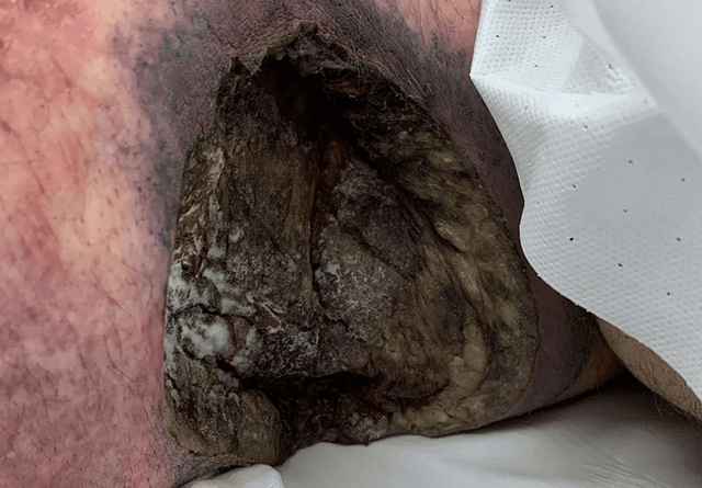 Left lateral chest wall with the apparent mold-like substance on the necrotic base
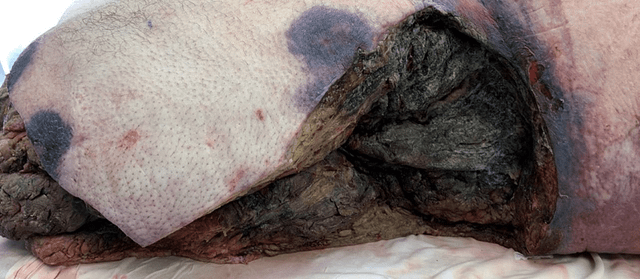 Left lateral thigh after above-knee amputation and multiple debridements with rapid progression of necrotic tissue
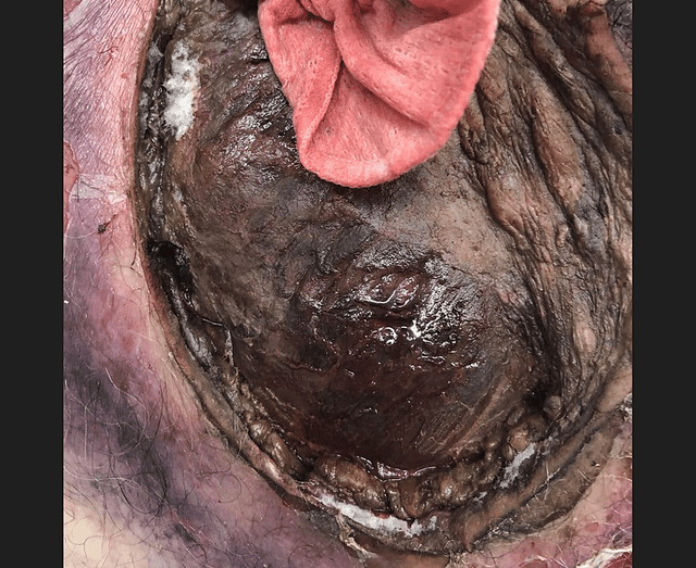 Left lateral chest wall approximately nine hours after prior debridement