

Cephalopagus is a rare variety of conjoined twins. They are fused with their heads, thoracic and upper abdominal cavities. The exact mechanism for development of conjoined twins cannot be clearly explained. It appears that there is an alteration in the normal developmental process of monozygotic twins, which fail to separate from each other. We present the morphology of a cephalothoracopagus, revealed through anatomical dissection, emphasizing the arrangement of the viscera in the thoracic and abdominal cavities. They are fused with their heads, thoracic and upper abdominal cavities. The lower abdomen and pelvic cavities are free. Each twin has two upper and lower limbs, normally shaped. Each twin has a heart and two lungs. There is a single pharynx, esophagus and stomach, but normal lower abdominal systems. The genital and urinary systems are apparently normal. Due to the fusion of the heads and abnormal arrangement of the superior central nervous system, surgery is not attempted in these cases, the prognosis being very poor.
Case Presentation:
We present the morphology of a cephalothoracopagus, revealed through anatomical dissection. The specimen was obtained from the Department of Obstetrics and Gynecology, Emergency University Hospital, Bucharest, Romania, with the signed agreement of the patient, fixed in formaldehyde and dissected under a magnifying glass. The external appearance of the specimen showed the twins with a single cephalic extremity and a single “face” (Figures 1 and 2).
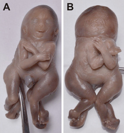
Fig1: External appearance of the specimen. Cephalothoracopagus deradelphus type. Duplication originating in the caudal region (dipygus [11])-two nearly complete components joined front to front, single neck and with heads more or less completely fused into a single compound mass. (A) Anterior view; (B) Posterior view; (C and D) Details. Note the clubfoot (blue arrows) (talipes equinovarus) of one of the "fetuses", the malformation involves the bones of the ankle and foot resulting in the adduction of the forefoot, inversion of the heel, and plantar flexion of the forefoot and ankle. Morphologically, there is a subluxation of the talocalcaneo-navicular joint. Because of this malformation, the dorsal aspect of the foot is often rotated medially, which assumes a clublike appearance.
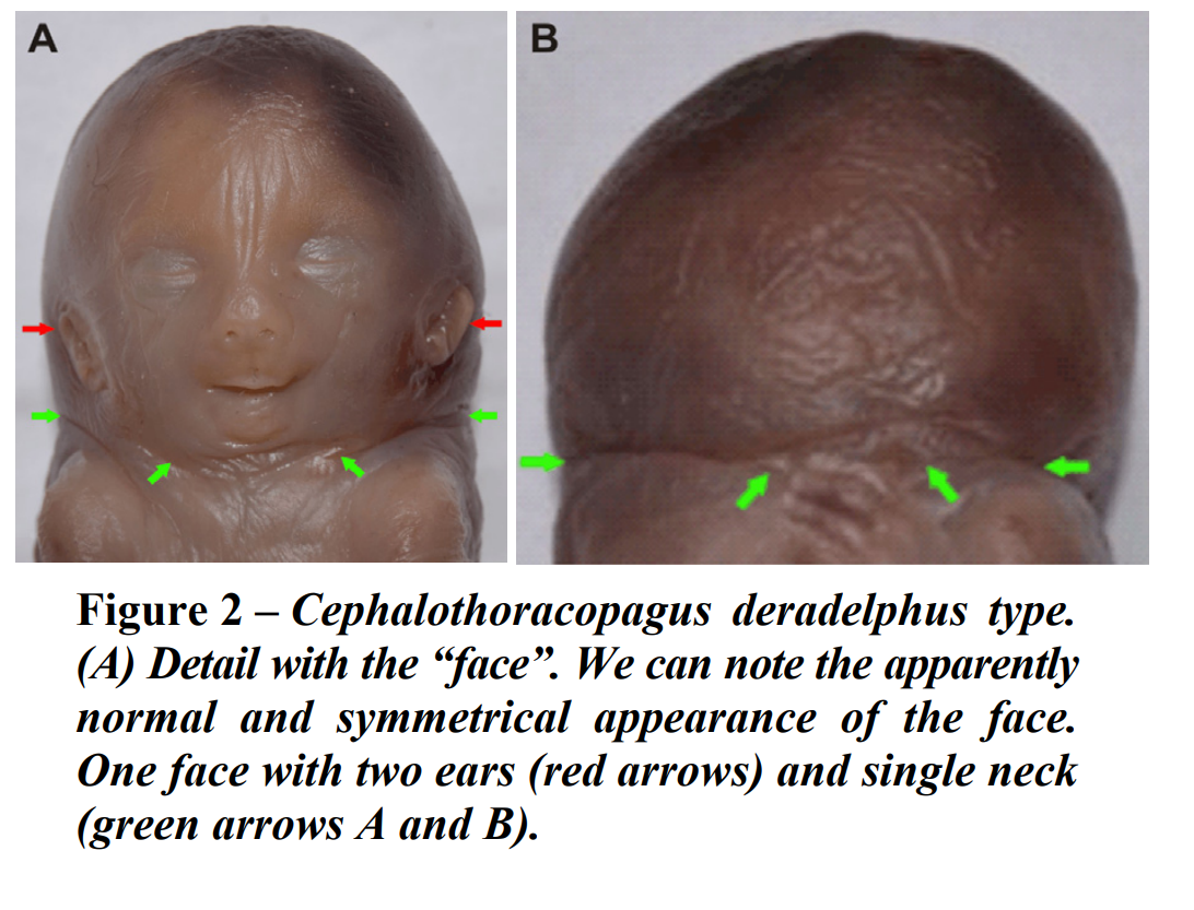
We considered the anterior surface the one with the “face”. The twins were joined at the head, the thoracic cavities and the upper abdominal walls. They were joined face-to-face and each had two upper and two lower limbs. The gender of the twins was masculine. We measured the specimen using standard calipers and the measurements showed a total height for both twins of 14 cm, measured from the vertex to the tip of the toes. The length of the thigh was of 3.3 cm, the calf 2.5 cm, the arm 2.4 cm and the forearm 2.3 cm. The transverse diameter of the head (the bi-parietal diameter) was 3.2 cm and the anterior–posterior diameter was 4.1 cm (measured from the nasion to the occipital protuberance). According to the measurements, we concluded that the gestational age was of about 15–16 weeks.
The cephalic extremity had apparently a single “face” with normal morphology. The extremity opposed to the face was slightly broader than normal, with two occipital protuberances. The dissection was performed under a magnifying glass. We performed an initial incision between the two twins on the median line, on the surface corresponding to the “face”, the anterior surface. The arrangement of the viscera in the abdominal and thoracic cavities was fairly complex, and difficult to assess due to their spatial disposition. The thoracic and abdominal cavities were separated through an apparently normal diaphragm (Figure 3).
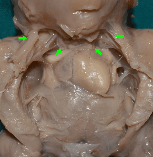
Fig3: – Cephalothoracopagus deradelphus type. Aspect of the arrangement of the viscera after first incision. Note the presence of one single neck (green arrows).
The thoracic cavity had a heart–lung complex for each of the twins. The “anterior” heart, according to our incision, was well developed and had four cavities. The “posterior” heart was less developed and its cavities could not be assessed (Figure 4).
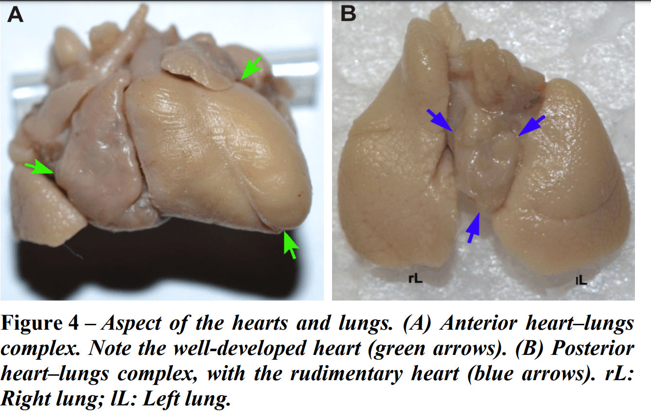
Each of the hearts had an apparently normal heart base, with normal vascular arrangement. From each heart branched an aorta with a normal pathway in the thoracic and abdominal cavities. All four lungs were slightly under-developed, but apparently normal in morphology. Each lung had a main bronchus and each pair of lungs was connected to a trachea. Between the two tracheas, we only found a single esophagus, following a common pharynx.
In the abdominal cavity, we found a larger “anterior” liver, and a smaller “posterior” one. A single under-developed stomach was discovered, following the esophagus between the two livers. Inferior to the stomach, the digestive tract divided into two intestines, which had an apparently normal pathway to the anus (Figure 5).
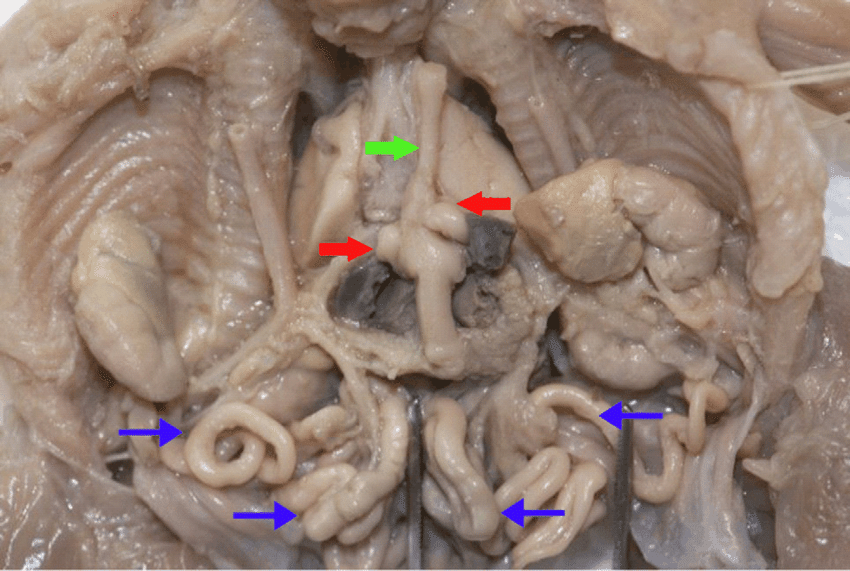
Fig5: – Aspect of the abdominal and thoracic cavities after removal of the anterior heart–lungs complex and liver. Note the single esophagus (green arrow) and stomach (red arrows) and also the two intestinal tracts (blue thin arrows).
The superior mesenteric artery of each digestive system branched from the ipsilateral aorta. The urinary tracts were normally developed with two kidneys for each twin, on each side of the vertebral column, followed by apparently normal ureters (Figure 6).
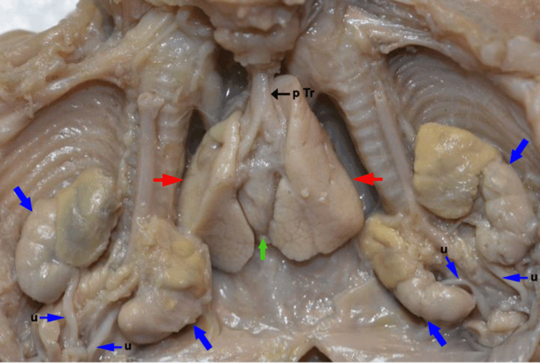
Fig6: Aspect of the abdominal and thoracic cavities after removal of the intestinal tract. Note the position of the posterior heart–lungs complex (red arrows – lungs and green arrow – heart) and the disposition of the urinary tracts (blue arrows). pTr: Posterior trachea; u: Ureter.
Discussion:
Conjoined twins are a rare complication of monozygotic twining. The earliest historic example of conjoined twins dates from the sixth millennium BC, in the form of a marble statuette portraying parapagus twins [2, 12]. The first documented case is the case of the Biddenden maids, in 1100 AD, who lived together for 34 years, joined at the hips and shoulders [2, 13]. The first successful separation of conjoined twins was performed in Basel by Johannes Fatio in 1689 [2]. Conjoined twins develop from a single fertilized ovum [2]. There are two common theories regarding development of conjoined twins: the fissure theory and the fusion theory. The fissure theory states that there is an incomplete separation of the embryonic discs in the 15th to 17th days of gestation. This theory however was contradicted by the fusion theory, advocated by Spencer and other authors, considering that conjoined twining cannot appear from a fission event. According to their theory, development of all various types of conjoined twins can be explained through the union between two distinct embryos taking place in the early embryonic stage [4–6, 14, 15]. The exact mechanism accounting for development of conjoined twins is not completely described. There are several varieties of conjoined twins, and several classifications, according to the site of fusion. There are also some classifications for symmetrical and asymmetrical types of conjoined twins. Some classifications include several more types resulting from the extension of the junction, like cephalo-thoracopagus, thoraco-omphalopagus and others [1]. In our case, we considered the classification proposed by Spencer in 1996, with eight major types, grouped according to their ventral or dorsal attachments [3, 7]. It is sometimes rather difficult to classify conjoined twins according to thestandard groups, due to their high variability. In our case, the specimen was cephalopagus variety according to the classification proposed by Spencer. The cephalopagus variety is extremely rare, and it is thought to appear one in 58 cases of conjoined twins [16]. Considering the general incidence of conjoined twins, we can conclude that this particular variety is indeed extremely rare. This particular variety has a fused head and often is fused at the level of the thorax and upper abdomen, has four arms, four legs, and separate lower abdomen and pelvis [3, 7].
This variety contains another smaller group called janiceps, named after the two-faced roman god Janus [17]. This latter group is composed of cephalopagus conjoined twins with two faces present, one on each side of the head. The specimen examined by us presented with a single apparently normal “face”, thus not a janiceps. The cephalic extremity had on one side an apparently normal “face”, and two occipital protuberances on the opposite side. Cephalopagus conjoined twins usually have four upper and four lower limbs, normal in appearance, as in the case presented by us. The anatomical analysis of cephalothoracopagus twins published by Baron et al., the limbs were normal in eight out of 10 cases [18]. The arrangement of thoracic viscera in cephalothoracopagus twins was described in a paper by Badawy & Shehata [10]. They described two hearts, one of them well developed and the other one much smaller and rudimentary in form. The same morphology was discovered in our case, with the “anterior” heart well developed and the “posterior” one, much smaller. We could only assess the cavities of the first one, and they were normal. We also found that the lungs were present for each of the twins, and had a normal morphology. The digestive system is interesting. The twins shared in our case a single pharynx and esophagus, as well as a single stomach, but this led on to two separate intestines with a normal pathway to the pelvic region.
The same arrangement is described by Badawy & Shehata [10]. Cephalopagus conjoined twins are usually non-viable due to numerous malformations especially involving the central nervous system. It is to be expected that the brain and spinal cords would be extremely abnormal, thus leaving no possibility for satisfactory surgical separation. In this particular variety of conjoined twins, most authorities do not recommend surgical intervention. In the majority of cases, this type of twins does not survive until birth, or die shortly afterwards [4]. Conclusions Cephalopagus is a rare variety of conjoined twins, representing about 11% of all conjoined twins. They are fused with their heads, thoracic and upper abdominal cavities. The lower abdomen and pelvic cavities are free. Each twin usually has two upper and lower limbs, normally shaped. There is a single pharynx, esophagus and stomach, but normal lower abdominal systems. The genital and urinary systems are apparently normal. The heart and lungs are present for each of the twins; usually, one of the hearts is less developed and rudimentary. Due to the fusion of the heads and abnormal arrangement of the superior central nervous system, surgery is not attempted in these cases, the prognosis being very poor.
Cîrstoiu, Monica-Mihaela & Filipoiu, Florin & Bratila, Elvira & Berceanu, Costin & Cirstoiu, Catalin & Budu, Vlad & Bulescu I., I. & Munteanu, Octavian. (2016). Morphological study of cephalothoracopagus deradelphus type conjoined twins. A case report. Romanian journal of morphology and embryology = Revue roumaine de morphologie et embryologie. 57. 249-252.